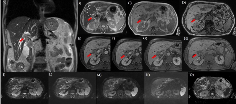
Fig 1. Fifty years old man with rectal cancer.
In A (T2-W HASTE coronal plane) and B (T2-W HASTE axial plane), the lesion (arrow) appears as a single periductal hyperintense mass extending to the bifurcations of both right and left hepatic ducts. In C (axial T1-W in phase) and D (axial T1-W out phase) the lesion (arrow) show hypointense signal. During arterial (E), portal (F), equilibrium (G) phase of contrast study the lesion has a progressive contrast enhancement while during hepatobiliary phase (H), is hypointense. DWI sequences: In I b0 s/mm2, in L b50 s/mm2, in M b400 s/mm2, in N b800 s/mm2 and in O ADC map. The tumor shows restricted diffusion.

