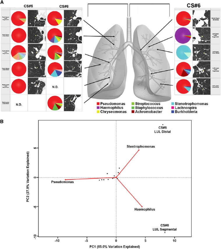
Figure 3.
(A) Bacterial communities present in individual lung airways. Multiple samples were taken from lung explants (right lung, subject CS#5; both lungs, subject CS#6) at the time of elective transplantation. Samples were harvested from the regions of lung indicated by the arrows on the gray lung schematic. Pie diagrams depict the genus level classification of 16S sequences, and the computed tomography images demonstrate the absence of bronchiectasis in the airways adjacent to where samples were obtained. The key for the nine most abundant organisms is provided below the lung schematic. (B) Cluster analysis of the bacterial communities sampled from sites in the left upper lobe (LUL). Biplot of the principle components analysis of the normalized bacterial communities from multiple anatomic sites in the lung explants. Reprinted by permission from Reference 73.

