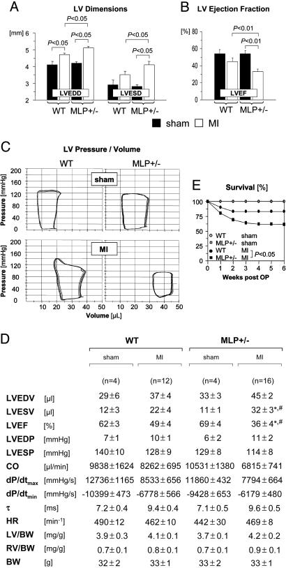
Fig. 2.
Enhanced LV dilatation, reduced systolic function, and increased mortality after MI in MLP+/– mice. (A and B) Echocardiography was performed 6 weeks after sham operation (filled bars) or MI (empty bars). Data are from n = 7 sham-operated mice per genotype, n = 18 infarcted WT mice, and n = 30 infarcted MLP+/– mice. LVEDD and LVESD denote LV end-diastolic and end-systolic diameters, respectively; LVEF, LV ejection fraction. (C and D) Pressure-volume loops were recorded 6 weeks after sham operation or MI. (C) Representative recordings are shown. (D) Data are summarized. LVEDV and LVESV denote LV end-diastolic and endsystolic volumes, respectively; LVEF, LV ejection fraction; LVEDP and LVESP, LV end-systolic and end-diastolic pressures, respectively; CO, cardiac output; dP/dtmax, maximal rate of pressure development; dP/dtmin, maximal rate of pressure decay; τ, monoexponential time constant of LV relaxation; HR, heart rate; LV, LV weight; RV, right ventricular weight; BW, body weight. *, P < 0.05 vs. sham-operated MLP+/– mice; #, P < 0.05 vs. infarcted WT mice. (E) Six-week mortality rates were assessed in 30 WT and 61 MLP+/– mice surviving for at least 48 h after coronary ligation. Sham-operated WT (n = 8) and MLP+/– mice (n = 7) served as controls (survival curves in sham-operated mice are superimposed).

