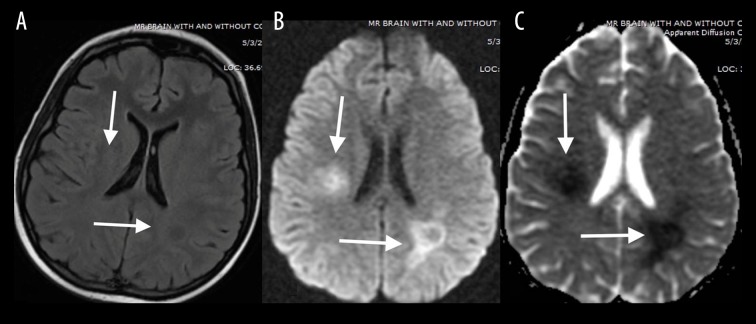
Figure 1.
A 15-year-old girl treated for acute lymphoblastic leukemia who presented with headache after methotrexate treatment initiation. Axial T2-FLAIR image (A) demonstrates slightly increased signal intensity with restricted diffusion involving periventricular white matter and centrum semiovale (white arrows) as seen on DWI (B) and ADC (C) images.

