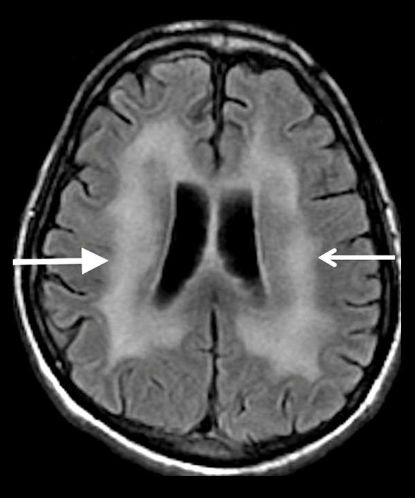
Figure 10.

A 40-year-old man with a history of AIDS. Axial T2-FLAIR image shows diffuse cerebral atrophy that is out of proportion to the patient’s age, and symmetrically increased T2 signal in the periventricular and deep white matter (white arrows) sparing the subcortical U-fibers.
