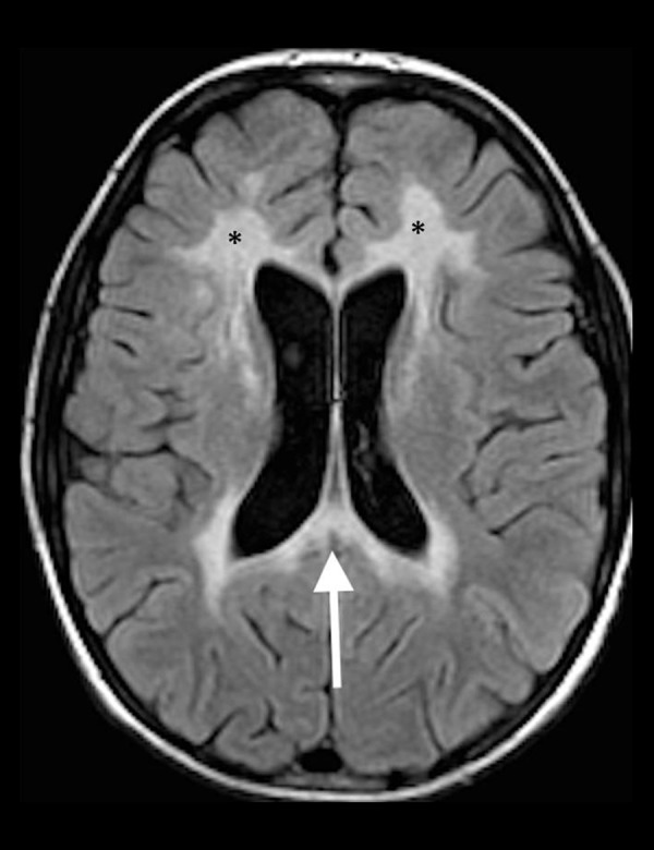
Figure 12.

A 14-year-old boy with X-linked adrenoleukodystrophy. Axial T2-FLAIR image shows increased T2 signal in the peritrigonal regions extending across the splenium of the corpus callosum (white arrow) as well as the involvement of the frontal lobe white matter bilaterally (black asterisks).
