
Figure 8.
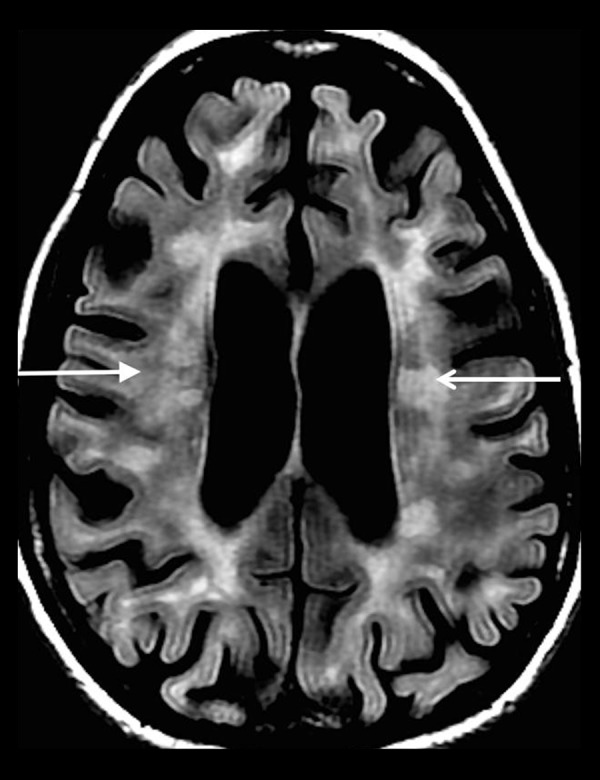
A 52-year-old woman with a long-standing history of multiple sclerosis. Axial T2-FLAIR image shows areas of increased T2-FLAIR signal in the bilateral periventricular white matter (white arrows) oriented perpendicular to the ventricular surfaces.