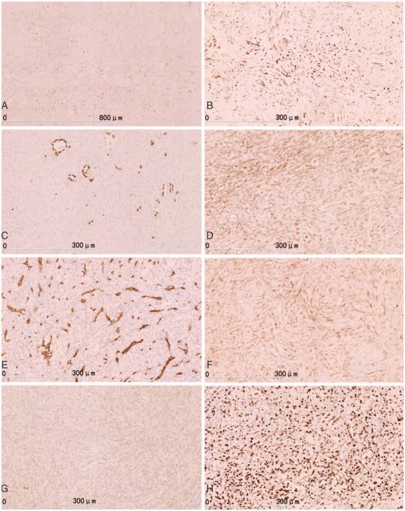
Figure 3.

Immunohistochemical staining. (A) CK (pan) showed tumor cells were positive. (B) CK7 showed positive in tumor cells. (C) TTF-1 was expressed in only type II alveolar epithelial cells, but tumors cells were negative. (D) Vimentin showed positive tumor cells. (E) CD34 showing vascular positive and tumor cells (−). (F) β-Catenin showed as cytoplasm positive in tumor cells, but were negative in tumor cells nuclei. (G) ALK showed tumor cells were negative. (H) Ki-67 showed a high tumor proliferative index (about 70%+). ALK = anaplastic lymphoma kinase, TTF-1 = thyroid transcription factor-1.
