Fig. 1.
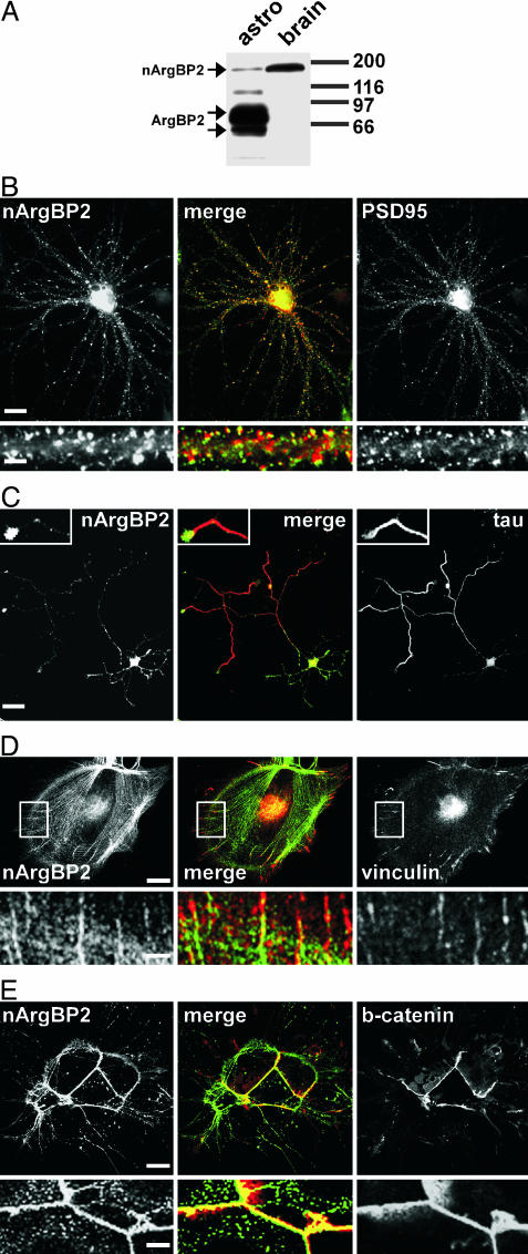
Expression of ArgBP2/nArgBP2 in brain and in primary cultures of brain cells. (A) ArgBP2/nArgBP2 immunoreactivity in total homogenates of astrocytes in primary culture and of brain tissue as revealed by Western blotting. nArgBP2 is the prevalent isoform in total brain, whereas the two ArgBP2 bands predominate in cultured astrocytes. (B and C) Localization of endogenous nArgBP2 in cultured hippocampal neurons. In mature (21 days in vitro) neurons (B), nArgBP2 is concentrated at synapses, primarily in dendritic spines, as revealed by double immunofluorescence for nArgBP2 and PSD95. A dendritic shaft is shown at higher magnification under each field. (Scale bars: 20 μm for the main fields; 5 μm for the small fields.) In developing isolated neurons (3–5 days in vitro)(C), nArgBP2 is localized throughout the dendritic arbor but is also present in axons (counterstained with the axonal marker tau) where it is concentrated in axon terminals. (Scale bars: 30 μm for the main field; 10 μm for the Insets.) (D and E) Localization of endogenous ArgBP2 in astrocytes. ArgBP2 is localized on stress fibers and at focal adhesions as demonstrated by counterstain with antibodies directed against vinculin, a focal adhesion marker (D). The portion enclosed by a white rectangle is shown at high magnification in Lower. (Scale bars: Upper, 20 μm; Lower, 4 μm.) In confluent cultures (E), ArgBP2 is concentrated at sites of cell–cell contact, where it colocalizes with β-catenin. (Scale bars: Upper, 10 μm; Lower, 2 μm.)

