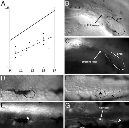
Fig. 1.
Peripheral growth of the efferent axons. (A) plot of the position of the leading efferent growth cones (ordinate) as a function of the developmental stage of the embryo (abscissa). The developmental stage is expressed as the position reached by the leading edge of the primordium, which is more accurately defined than its trailing edge (15). Both the position reached by the leading efferent neurite (ordinate) and the position reached by the leading edge of the primordium (abscissa) are expressed as somite number. Because the primordium extends over 2.5 somites on average, the diagonal line represents the average position of the trailing edge of the primordium. In this sample of 19 embryos, the leading efferent neurite lags anywhere between 3 and 9 somites behind the trailing edge of the primordium. The dashed line is the least-squares regression line for the positions of the efferent leading edges. (B and C) In a 32 hours after fertilization (haf) sdf1a morphant embryo, where the primordium fails to migrate (B), the efferent fiber reaches the trailing region of the primordium and stops there (C). When the primordium eventually differentiates into one or two ectopic neuromasts, these are invariably innervated by efferent neurites (not shown). (D–G) Efferent innervation in 3-day-old Zath1 morphant embryos. (D and E) A neuromast that comprises no hair cells (dotted outline) is nevertheless contacted by an efferent fiber (arrowhead). (F and G) Two terminal neuromasts, one of which (asterisk) comprises a hair cell and the other not. The neuromast with no hair cell is contacted by an efferent fiber (arrowhead). (Final magnification: B and C, ×150; D–G, ×130.)

