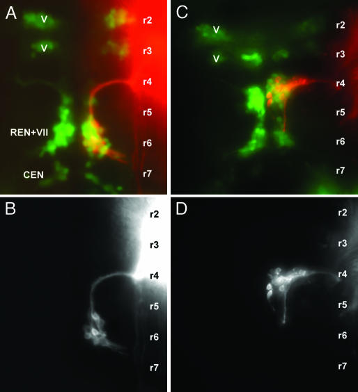
Fig. 5.
Central pathways of facial motor neurons in 2-day-old wild type (A and B) and sdf1a morphant embryo (C and D). The facial neurons were labeled with DiI and are shown either alone (B and D) or in an islet-GFP background (A and C; red, DiI; green, GFP). The approximate localization of the rhombomeres (r1, r2,...) was defined as in Fig. 3. V, trigeminal motor nucleus; VII, facial motor nucleus; REN, rostral efferent nucleus of the lateral line system; CEN, caudal efferent nucleus of the lateral line system.

