Fig. 3.
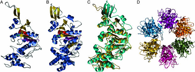
Homology model for the C terminus of VirB4. (A) VirB4 monomer model. α-helices are blue and β-sheets are gold. The Walker A motif (G433-T440) is red, the Walker B motif (R619-E635) is magenta, and the conserved Q668 is green. (B) Monomer of TrwB, using the same coloring as for VirB4. (C) Superposition of VIRB4 (turquoise) and TrwB (gold). (D) Predicted hexameric structure of VirB4.

