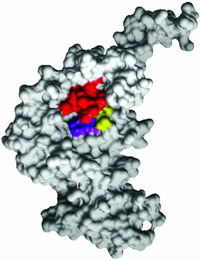
Fig. 5.
Surface image of VirB4 monomer, highlighting the NTP-binding cleft. To better display the NTP-binding cleft, we show the structure from a different perspective, corresponding to the back side of Fig. 3A. The conserved lysine (K) of the Walker A motif is red, the conserved DE of the Walker B motif is purple, and the conserved glutamine (Q) is highlighted in yellow.

