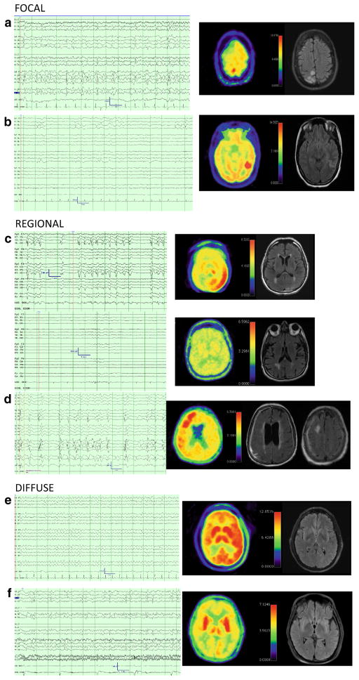
Fig. 1.
Representative cases of patterns of PET hypermetabolism with corresponding electrographic and MR data. 13-s cEEG samples and FDG-PET imaging [rainbow color scheme; 10 % gradations from hypometabolic (blue) through green and yellow to hypermetabolic (red), cerebellum set to yellow, L demarcates left orientation] and FLAIR MR images for six subjects. a, b are representative cases of focal hypermetabolism. a (SUBJ 3) is a 32-year-old woman with a history of peri-natal stroke that presented with epilepsia partialis continua, MRI had T2 hyperintensity near the remote infarct with corresponding PET hypermetabolism in the same region. EEG had LPDs and intermittent discrete seizures. She required surgery to remove the ictal focus, which resulted in cessation of seizure activity, but a worsened hemiparesis. b (SUBJ 1) Is a 25-year-old woman that presented with convulsive status epilepticus transitioning to persistent encephalopathy with initial AED treatment. cEEG had persistent 0.5 Hz right posterior quadrant LPDs, MRI revealed a posterior temporal T2 hyperintensity with an adjacent developmental venous anomaly. PET revealed focal hypermetabolism in the region of the MRI abnormality. Treatment was escalated with IV sedation and patient made a full recovery. Follow-up MRI revealed a resolution of T2 hyperintensity. c, d are representative cases of regional hypermetabolism. c (SUBJ 15) is a 77-year-old man presenting with hypertension and T2 hyperintensities of the left occipital/parietal cortex and scattered white matter T2 hyperintensities, DWI changes, and microhemorrhages. Initial cEEG revealed discrete electrographic seizures. After anti-seizure medication (top panel), cEEG displayed an IIC pattern (left LPDs) associated with focal FDG-PET hypermetabolism in left parietal, occipital, posterior-frontal right thalamic regions. (Bottom panel) Anesthetic burst-suppression was employed and repeat PET yielded resolution of hypermetabolism 12 days later concurrent with burst-suppression on cEEG. LPDs subsequently resumed with weaning sedation and his persistent encephalopathy prompted family to elect palliative care, autopsy revealed inflammatory cerebral amyloid angiopathy. d (SUBJ 13) is an 84-year-old woman with dementia s/p VP shunt placement for NPH that developed discrete focal seizures, which resolved with AED treatment. Patient had persistent encephalopathy and was found to have LPDs on cEEG and regional hypermetabolism on PET. MRI revealed pachymeningeal thickening and enhancement as well as a right frontal focal area of hemorrhage and surrounding T2 hypermetabolism along the shunt tract. Patient did not improve and was transitioned to palliative care. e, f are representative cases of diffuse hypermetabolism. e (SUBJ 11) is 44-year-old man with subacute cognitive decline presenting with clinical seizures and worsening encephalopathy. cEEG had 4 Hz GRDA and diffuse (L > R) cortical and subcortical hypermetabolism. Found to have VGKC antibody, return to baseline with AEDs and IVIG. f (SUJB 5) is a 23-year-old man presenting with NORSE, his clinical seizures improved with treatment, but clinical deficits persisted. cEEG revealed LPDs. PET has bilateral hypermetabolism primarily in the basal nuclei. MRI was normal other than mild generalized volume loss. Patient recovered with a residual modest spastic tetraparesis and cognitive recovery. Etiology of illness remains cryptogenic (Color figure online)

