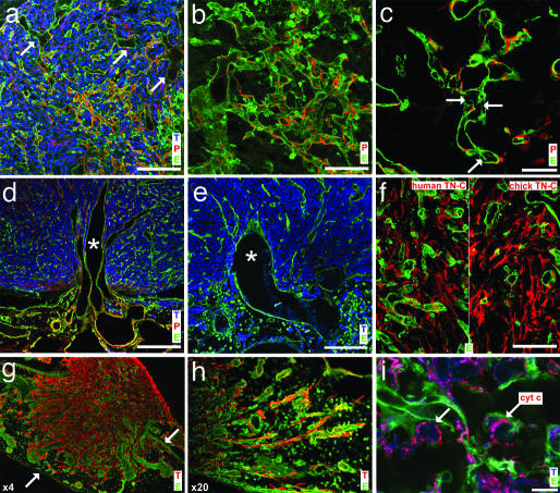
Fig. 3.
Immunohistological and confocal microscopy analysis of experimental glioma. (a) Frozen sections of D7 tumors were stained with antivimentin antibody (tumor cells, T), antidesmin antibody (pericytes, P), and fluoresceincoupled SNA-1 lectin (endothelial cells, E). Note the high tumor cell density, the irregular and dilated vessels (arrows), and pericyte coating of most capillaries. (b) Tumor capillaries form glomeruloid-like microvascular proliferations, covered by pericytes. (c) The presence of intercapillary pillars (arrows) in tumor capillaries shows intussusceptive angiogenesis. (d) Cooption of larger, dilated CAM blood vessels at the base of the tumor. (e) Flexion and orientation of a coopted vessel toward the tumor. (f) The presence of TN-C in the extracellular matrix of glioma identified by species-specific mAbs. (g) Lowpower magnification illustrates the invasive behavior of the tumor (arrows mark the borders of the CAM). (h) Individual glioma cells invade the CAM adjacent to the implantation site and migrate along blood vessels. (i) Experimental glioma do not exhibit apoptosis as revealed by physiological distribution of cytochrome c around cell nuclei (arrows). (Scale bars: a, d, and e, 100 μm; b and f, 50 μm; c, 20 μm; and i, 10 μm.)

