Fig. 4.
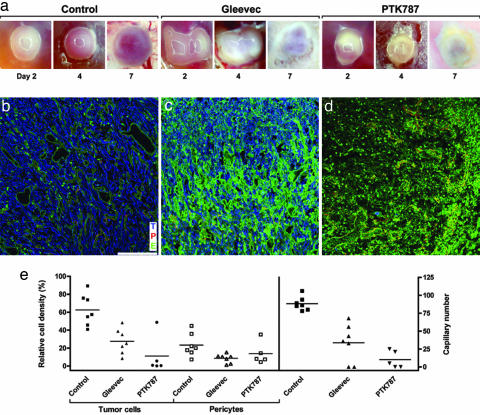
Topical treatment of experimental glioma with receptor tyrosine kinase inhibitors Gleevec and PTK787/ZK 222584. (a) Biomicroscopy follow-up of control and treated tumors. Control tumors show progressive growth and vascularization, whereas Gleevec- and PTK787/ZK 222584-treated tumors appear white and show signs of tissue damage. (b–d) Confocal microscopy analysis of triple-stained tumors at D7. Tumor cells (T) are blue, pericytes (P) are red, and the endothelium (E) is green. (b) Control tumors show densely packed tumor cells within a well established pericyte-covered vascular network. (c) Gleevec-treated tumors show a strong decrease in tumor cell, pericyte, and vessel density. The increase in green fluorescence is caused by nonspecific lectin binding to necrotic cells. (d) In PTK787/ZK 222584-treated tumors, only a few pericyte-covered capillaries are found after treatment, and almost all tumor cells have disappeared. As in Gleevec-treated tumors, nonspecific green fluorescence increases. (e) Quantification of receptor tyrosine kinase inhibitor effects on glioma development. Bars indicate means of relative cell density and vessels counts. (Magnification: a, ×10; scale bar: b–d, 100 μm.)

