FIGURE 4.

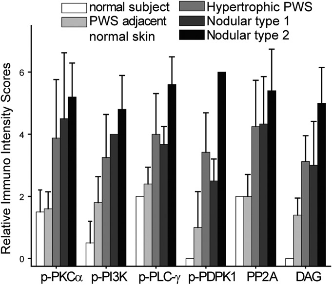
Relative IR intensity scores of p-PI3K, p-PKCα, p-PLC-γ, p-PDPK1, DAG, and PP2A in the blood vessels from different types of PWS lesions.

Relative IR intensity scores of p-PI3K, p-PKCα, p-PLC-γ, p-PDPK1, DAG, and PP2A in the blood vessels from different types of PWS lesions.