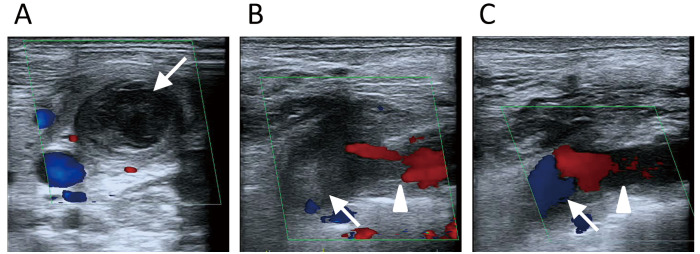
Figure 1.
Ultrasonography images (color flow Doppler imaging). A: A short-axis view of the left side of the neck. On the first ultrasound examination, a hyperechoic region of the thrombus in the left jugular vein thrombus coexisted with a hypoechoic region, which occluded the left jugular vein (arrow). B: The left subclavian approach. On the first ultrasound, a thrombus was present in the left subclavian vein (triangle) and another thrombus was present in the left brachiocephalic vein (arrow). C: The left subclavian approach. After 1 month of edoxaban treatment, the thrombi in the left subclavian vein (triangle) and left brachiocephalic vein (arrow) were smaller, and blood flow through these veins was increased.

