Figure 1.

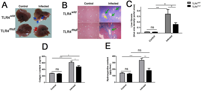
The mice with TLR4 mutation showed an attenuated pathogen-associated biliary fibrosis caused by Clonorchis sinensis. The mice with TLR4 wild and mutation were orally administrated by 45 metacercariae C. sinensis or PBS, respectively. These mice as well as healthy control mice were executed and livers were harvested on 28 days of C. sinensis post-infection. (A) Gross clinic hepatic changes of TLR4wild and TLR4mut C3H mice caused by C. sinensis. Blue arrows indicate massive nodules with different size in the hepatic lobes in the TLR4wild mice infected by C. sinensis, and red arrows showed limited pale area on the surface of lobes without nodules in TLR4mutmice, compared with TLR4wild mice. (B) Representative Masson staining slides showed collagen fiber deposition (blue stripes). Scale bars = 200 μm. Green arrows showed the proliferation of biliary epitheliums and enlarged the bile ducts in the mice of C. sinensis-infected mice, and the yellow arrow shows the worm’s body in the enlarged bile ducts. (C) The mean optical density of collagen fibers indicated by Masson staining was digitized and quantitated in the liver of healthy and C. sinensis-infected mice by on Image-Pro Plus software. (D) Concentrations of Collagen I in the hepatic homogenate of healthy control and C. sinensis-infected mice with TLR4wild and TLR4mut. (E) Concentrations of hydroxyproline in the hepatic homogenate of healthy normal control and C. sinensis-infected mice with TLR4wild and TLR4mut. The data were obtained from 3~5 mice of three-independent experiment. The values were expressed as mean ± SEM. compared with indicated group, *P < 0.05, **P < 0.01, ***P < 0.0001.
