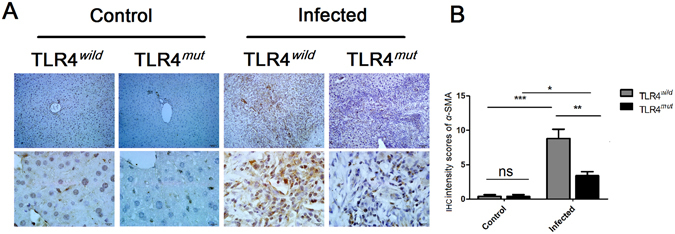
Figure 5.

Impairment of TLR4 attenuates the activation of HSCs in the PABF caused by Clonorchis sinensis. Livers from TLR4 wild and mutated mice infected with or without C. sinensis were harvested, and the sections of tissue containing bile ducts were fixed by 4% formalin solution for IHC. The activation of HSCs was indicated by immunohistochemical staining of α-SMA. (A) Representative images of α-SMA staining on HSCs of TLR4 wild or mutated mice as indicated. Scale bars for upper panel are 50 μm and for the nether panel are 10 μm, respectively. (B) The expression of α-SMA were semi-quantitated by independent pathologists as described in Methods and Materials. The data were obtained from 3~5 mice of three-independent experiment. The values were expressed as mean ± SEM. Compared with indicated mice, *P < 0.05, **P < 0.01, ***P < 0.001.
