
Fig. 4.
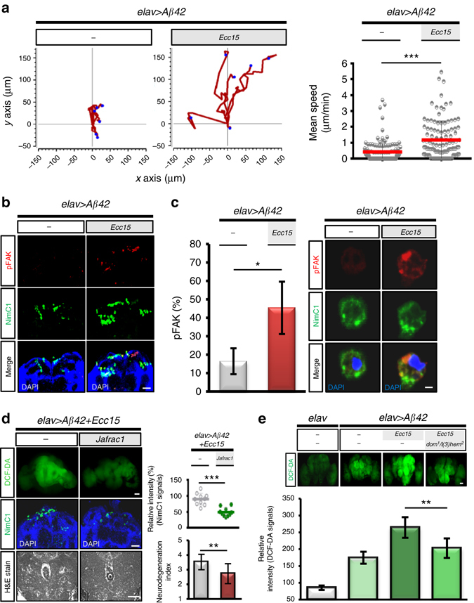
Gut–brain axis mediates the mobilization of hemocytes and their attraction to the transgenic brain in promoting neurodegeneration. a Time-lapse analysis of plasmatocyte migration after enteric infection. Representative examples of the migration trajectories of plasmatocytes (n = 6 in each group). b, c Coimmunostaining of phospho-FAK and NimC1 in plasmatocytes recruited transgenic brains b or in circulation c. Phospho-FAK, red; plasmatocytes, green (n = 15 in each group). d DCF-DA (upper panel) and NimC1 (middle panel) staining and histology (bottom panel) in the transgenic brains with or without overexpressing Jafrac1. e DCF-DA staining in transgenic brains with or without hemocyte deficient background. elav > Aβ42 transgenic or control (elav alone) flies with or without Ecc15 intestinal infection were analyzed at 10 dpi. DCF-DA signal, ROS stress; NimC1 signal, plasmatocyte. Brain histology, n = 10 each group. Quantitative data are presented as the mean±SD of three independent experiments. *P < 0.05, **P < 0.01, ***P < 0.001; NS, not significant, H&E, haematoxylin and eosin. Scale bars, 50 μm b, d and e; 5 μm c