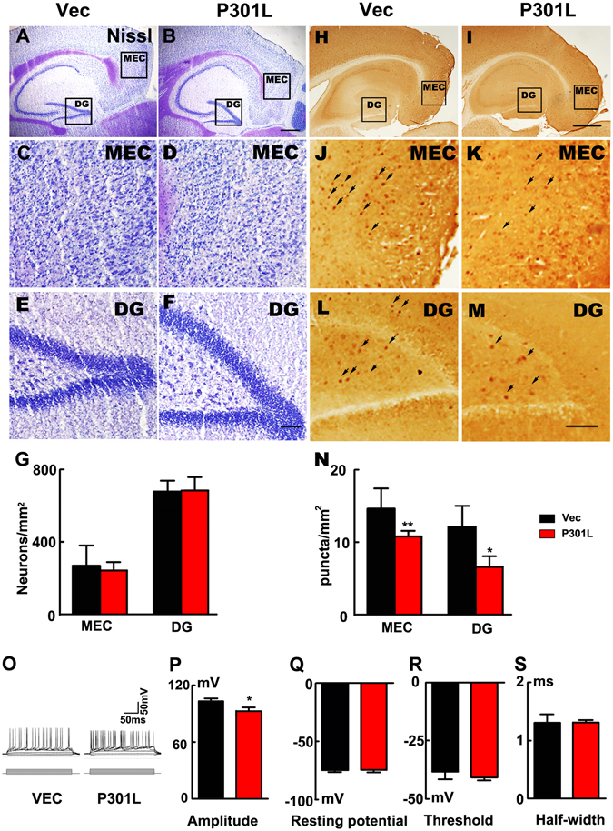
Figure 4.

Expression of P301L hTau in MEC suppresses the neuronal activity without causing significant cell death. (A–F) Nissl staining indicated that the numbers of neurons were not changed in P301L hTau transduced mice in both MEC and DG regions compared to control (scale bar in A and B, 500 μm; in C-F, 100 μm). (G) Quantitative analysis of the Nissl staining showed that P301L hTau transduction didn’t lead to neuronal death (n = 3 per group). (H–M) c-fos was employed to detect the neuronal activity. A density of the c-fos puncta in both MEC and DG subsets was remarkably decreased in P301L hTau-transduced mice compared with the control. (scale bar in H and I, 500 μm; in J-M, 100 μm). (N) Quantification of the number of c-fos positive cells per 1 mm2 revealed that P301L hTau lowered the c-fos expression in the MEC and DG. (n = 3 per group, 9 slices, *p < 0.05, **p < 0.01). (O–S) Whole-cell patch results suggested that AP amplitude was significantly lowered in P301L hTau cells compared to control (O,P). However, passive properties were not altered in P301L hTau cells since their membrane resting potential (Q) was unchanged compared to controls. There was no difference in threshold (R) or half width (duration, (S)) between control and P301L hTau cells (n = 3 per group, 9 slices, *p < 0.05). Arrows in J-M were used to indicate the cFos staining. Data were expressed as mean ± SEM.
