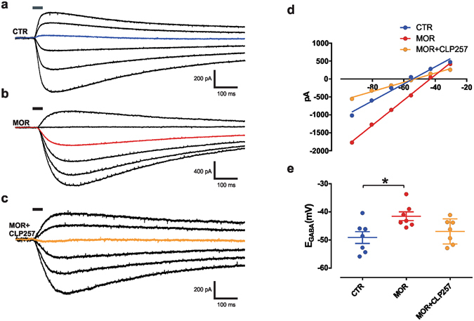
Figure 1.

Acute CLP257 restores Cl− extrusion in SDH neurons of morphine-treated rats. (a–c) Voltage clamp responses at different holding potentials to 30-ms muscimol puffs (solid line) in SDH neurons following saline (a) or morphine (b) treatments in the presence of a Cl− load (29 mM) to measure Cl− extrusion capacity. In (c), muscimol response in a SDH neuron from a morphine-treated rat obtained after slice pre-incubation with CLP257 (100 µM) for 1 hour. The colored line indicates the response at −55.5 mV (note the change in polarity). (d) I-V relationships for GABAA currents obtained from neurons in (a–c). (e) Pooled E GABA in controls (n = 7 cells, blue), in morphine-treated rats (n = 7 cells, red) and following CLP257 treatment (n = 7 cells, orange). E GABA is significantly more depolarized following morphine treatment as compared to control rats (one-way ANOVA, P = 0.02, Tukey post-hoc *P = 0.02), but not after CLP257 pre-incubation (P = 0.7); MOR vs. MOR + CLP (P = 0.1). Abbreviations: CTR = control; MOR = morphine; SDH = superficial dorsal horn.
