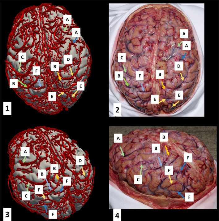
Figure 2.
Superficial vein segmentation results for Cadaver 1 overlaid on T1-weighted images. The left column (quadrants 1 and 3) shows the 3D reconstruction of the segmented veins overlaid on the brain surface. The corresponding photographs are shown in quadrants 2 and 4. Vein labels on the left correspond to those of the right in the same row. The key is as follows: A, frontal veins; B, parietal veins; C, left superior anastomotic vein (of Trolard); D, right superior anastomotic vein (of Trolard); E, right occipital veins; F, left occipital veins.

