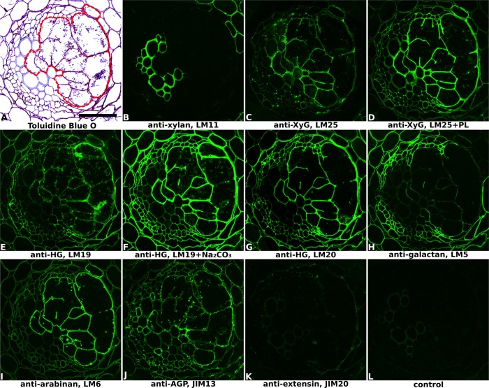
FIGURE 1.
Immuno-fluorescence imaging of syncytia induced by potato cyst nematode Globodera pallida within potato roots (cv. Desiree, 14 dpi). (A) The extent of the syncytium is indicated in the Toluidine Blue O stained bright field image with a red line. Indirect immunofluorescence (green) resulting from the binding of specific mAbs is shown for corresponding serial sections: (B) LM11 to heteroxylan; (C,D) LM25 to xyloglucan (XyG); (E,F) LM19 to non/low methyl-esterified homogalacturonan (HG); (G) LM20 to methyl-esterified HG; (H) LM5 to pectic galactan; (I) LM6 to pectic arabinan; (J) JIM13 to AGPs; (K) JIM20 to extensin. LM11 binds only to the xylem vessels in the vascular cylinder (B) so serves to identify these cells in all sections. Control section (L) was processed without primary antibody. PL, pre-treated with pectate lyase; Na2CO3, pre-treated with Na2CO3; Scale bar = 50 μm.

