Figure 3.

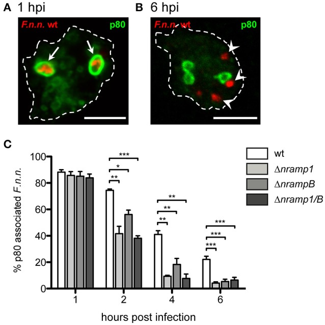
Phagosomal escape of F.n.n. in nramp knockout cells. (A,B) Representative micrographs of p80-positive and –negative F.n.n. in Dictyostelium wt cells at 1 and 6 hpi, respectively. Arrows indicate p80-positive F.n.n., arrowheads indicate p80-negative F.n.n. (C) Dictyostelium wt, Δnramp1, ΔnrampB, and Δnramp1/B cells were infected with F.n.n. wt GFP and the association of the endosomal marker p80 with the FP was monitored over 6 hpi by fluorescence microscopy (mean ± SEM, n = 3–6). Statistical analysis was performed applying a repeated measures one-way ANOVA analysis with Dunnett's post-hoc test for each timepoint: *p < 0.05, **p < 0.01, ***p < 0.001.
