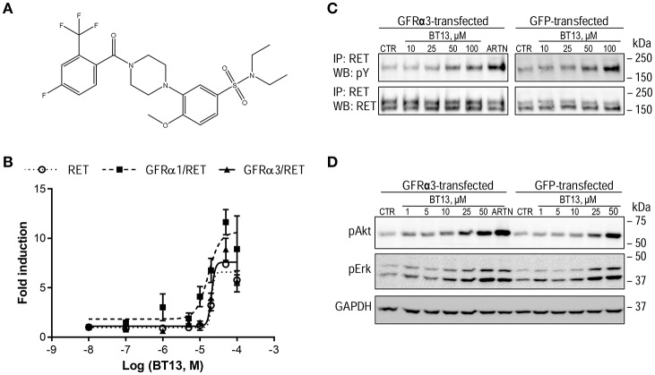
Figure 1.
The structure and biological activity of a small molecular weight RET agonist BT13. (A) Structure of BT13. (B) Fold induction of luciferase activity by BT13 in reporter cell lines expressing GFRα1/RET (GFRα1/RET, dashed line, black squares), GFRα3/RET (GFRα3/RET, solid line, black triangles), or RET (RET, dotted line, empty circles). (C,D) Activation of RET phosphorylation (C) and RET-dependent intracellular signaling (D) by BT13 in cells overexpressing GFRα3/RET (GFRα3-transfected) and GFP/RET (GFP-transfected). Luciferase activity was measured in three independent experiments for each concentration of BT13. GFRα—glycosylphosphatidylinositol (GPI)-anchored GDNF family receptor α; RET, rearranged during transfection; ARTN, artemin; IP, immunoprecipitation; WB, western blotting; pY, phosphotyrosine; pAkt, phosphorylated form of Akt protein; pErk, phosphorylated form of Erk1/2; GAPDH, Glyceraldehyde 3-phosphate dehydrogenase, house-keeping protein, loading control.

