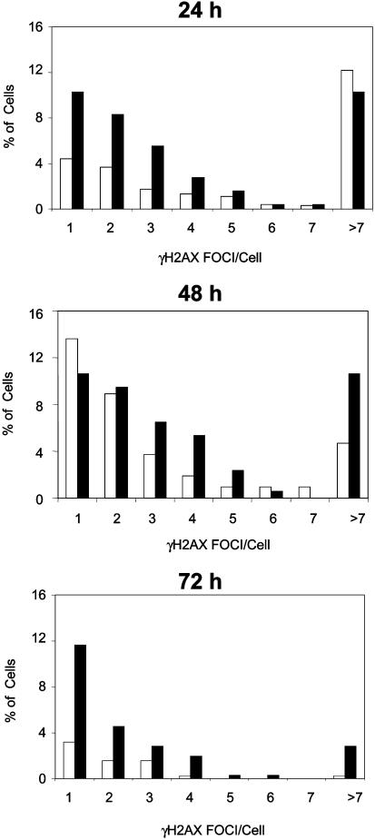
Fig. 4.
Generation and maintenance of γ-H2AX foci in conditionally Nbn-inactivated, activated B lymphocytes. γ-H2AX foci were stained with a specific antibody in CD19-Cre Nbnlox6/lox6 (black bars) and CD19-Cre Nbnlox6/WT B lymphocytes (white bars) after 24, 48, and 72 h of LPS/IL-4 activation. The samples were double-blinded and foci were scored by fluorescence microscopy. (WT/24 h, 976 cells; WT/48 h, 213 cells; WT/72 h, 379 cells; lox6/24 h, 253 cells; lox6/48 h, 169 cells; lox6/72 h, 352 cells.)

