Figure 2.
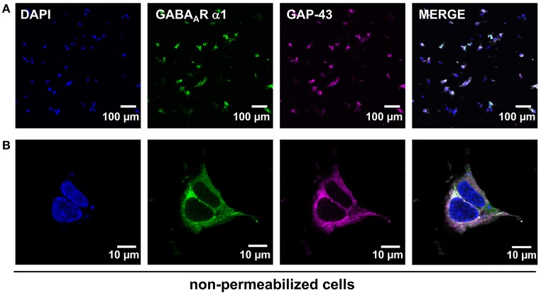
HEK293 transiently expressing GABAA α1β2 receptors and GAP-43 fused with red fluorescent protein. (A) Upper lane demonstrates transfection efficiency controlled by co-transfection of the membrane marker protein GAP-43 fused to dsRed (magenta). The α1 subunit of the GABAA receptor was stained specifically at the cellular surface (green). The merged picture represents co-localization resulting in a white signal of both the GABAA α1 receptor subunit and GAP43 (magenta) at the plasma membrane. (B) The lower lane shows an enlarged image of a single stained cell. DAPI staining was used to mark the nucleus of the cell (blue), α1 subunit is marked in green and GAP-43 in magenta.

