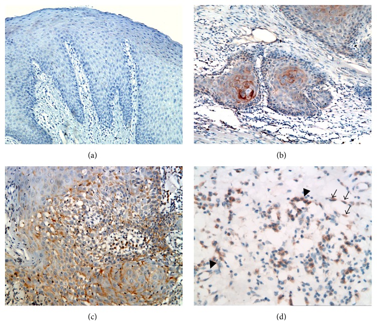
Figure 2.
Photomicrographs showing negative immunoreaction of VEGF-C in normal mucosa ((a) ×200), weak positive VEGF-C expression in node-free OSCC ((b) ×200), and more diffuse positive expression in positive lymph nodes OSCC ((c) ×200), ((d) ×400). (d) shows VEGF-C expression by cancer associated fibroblasts (arrows) and blood endothelial cells (arrow heads).

