
FIG 3 .
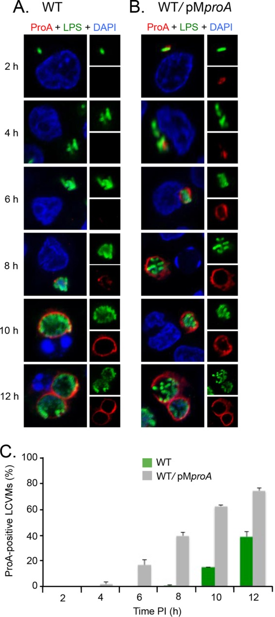
Timing of ProA localization to the LCVM. (A and B) U937 cells were infected for 2, 4, 6, 8, 10, and 12 h with either WT 130b (A) or the WT carrying pMproA (B) and were then labeled with LPS antisera (upper-right boxes) and ProA antisera (lower-right boxes) and analyzed by confocal microscopy. Host and bacterial DNAs were stained with DAPI, and the merged images appear as the larger boxes on the left side. Results presented here show a portion of the cell containing the LCV. (C) Quantification of percent ProA localization (± SD) to the LCVM over time, based on combined results from three independent trials.