
FIG 7 .
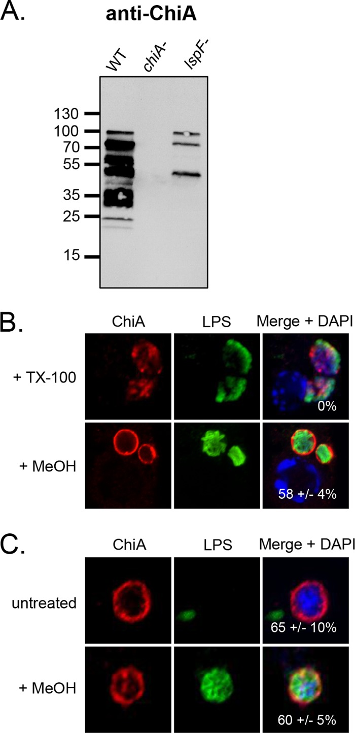
Location of ChiA in L. pneumophila-infected macrophages. (A) WT 130b, chiA mutant NU318 (chiA-), and lspF mutant NU275 (lspF-) were grown in BYE broth, and culture supernatants were then subjected to SDS-PAGE and immunoblot analysis with ChiA antiserum. (B) U937 cells were infected with WT 130b and permeabilized with TX-100 or MeOH and were then analyzed by confocal microscopy with ChiA (left column) and LPS (center column) antisera. The images presented show a portion of the cell containing the LCV. (C) LCVs obtained from WT 130b-infected U937 cells were labeled with ChiA and LPS antisera and then analyzed by confocal microscopy. (B and C) Host nuclei and bacterial DNAs were stained with DAPI, and merged images appear in the right columns with the percentages of ChiA-positive LCVMs (± SD) in the lower-right-hand corner. The data presented are representative of three independent experiments.