Fig. 4.
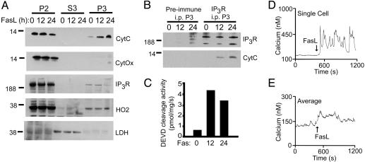
Extrinsic apoptotic signaling via FasL is associated with cytochrome c binding to IP3R and calcium mobilization. (A) Jurkat cells were treated with 1 ng/ml FasL vesicles for 0, 12, or 24 h, and the subcellular distribution of cytochrome c (CytC), cytochrome c oxidase (CytOx), IP3R, heme oxygenase-2 (HO2), and lactate dehydrogenase (LDH) was monitored in the mitochondrial-enriched 10,000 × g pellet (P2), the 100,000 × g cytosol (S3), or the light membrane-enriched 100,000 × g pellet (P3). (B) Immunoprecipitation of IP3R from P3 fraction solubilized in buffer A with anti-IP3R or preimmune serum. Samples were immunoblotted for IP3R and cytochrome c. (C) DEVD cleavage activity of the S3 fraction. (D and E) Calcium mobilization in response to FasL examined in individual fura 2-acetoxymethyl ester-loaded Jurkat cells. A representative single cell (D) and 50-cell average (E) from one experiment are shown. Approximately 30% of cells responded with an immediate and oscillatory rise in cytoplasmic calcium in response to FasL. All data are from the same experiment and are representative of at least three separate determinations.

