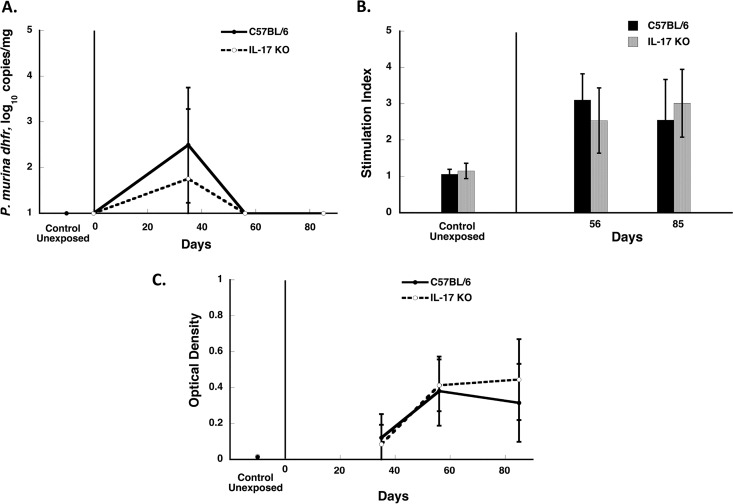
FIG 6.
Clearance of Pneumocystis infection in IL-17A KO mice. IL-17A KO and C57BL/6 mice were cohoused with a seeder mouse, and animals from each group were sacrificed at the indicated time points (days 35, days 56 to 58, and days 84 to 86 after the start of cohousing with an infected seeder). The results represent the means and standard deviations for mice from all cages within a single experiment at a given time point. (A) Pneumocystis organism load (as log10 dhfr copies/mg of lung tissue); (B) proliferative responses (indicated as the stimulation index) to crude Pneumocystis antigens; (C) anti-Pneumocystis antibodies (presented as the optical density) detected by ELISA using a crude Pneumocystis antigen preparation. Pneumocystis infection in the lung tissue was quantitated by Q-PCR using the single-copy dhfr gene as the target. There were no significant differences between C57BL/6 and IL-17A KO mice at any time point for any of the parameters.

