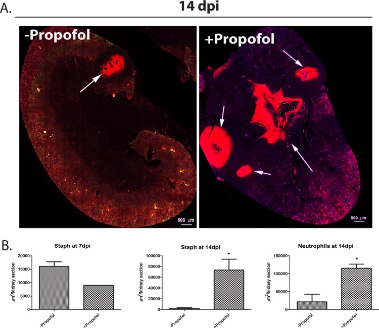
FIG 3.
Neutrophilic abscesses are not confined to the kidney cortex in propofol-treated mice. (A) Kidney sections from animals were obtained at 14 days postinfection, as described in the legend to Fig. 2. Sections were stained with antibodies against neutrophils (anti-Ly-6G; red) and with DAPI (blue). Propofol treatment induces formation of multiple abscesses in the kidney cortex as well as the central medullary region. Arrows indicate neutrophilic abscesses. Images were taken at 0.6× magnification; scale bar, 500 μm. Images are representative from 2 independent experiments with 5 animals per treatment group. (B) Quantitation of area of kidney section staining positive for presence of S. aureus or neutrophils at 7 days postinfection (top) and 14 days postinfection (bottom). *, P < 0.05.

