FIG 3.
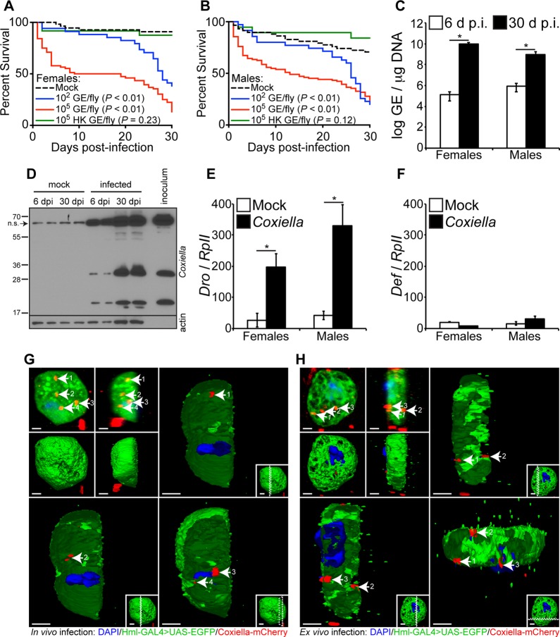
Adult Drosophila flies are susceptible to C. burnetii and elicit a host immune response. (A and B) Four-day-old Oregon-R female (A) and male (B) Drosophila flies were infected with live (102 or 105 GE/fly) or HK (105 GE/fly) C. burnetii, and survival was evaluated for 30 days. (C) Four-day-old adult Oregon-R flies were infected with C. burnetii (100 GE/fly), and bacterial levels were determined at 6 and 30 days postinfection by quantitative real-time PCR. (D) C. burnetii antigens were detected in infected flies at 6 and 30 days postinfection, as shown by immunoblotting using a rabbit polyclonal antibody against Coxiella. Biological duplicates are shown. Nonspecific (n.s.) banding from fly homogenates is denoted by the arrow. (E and F) Antimicrobial peptide levels of Drosocin (E) and Defensin (F) were determined in Oregon-R adults infected with C. burnetii (100 GE/fly) at 12 days postinfection. (G and H) Confocal microscopy showing mCherry-Coxiella invasion of hemocytes (white arrows) derived from 3rd-instar larvae infected in vivo (G) or ex vivo (H). The hemocytes expressed GFP, and the nuclei were stained with DAPI. Bars = 2 μm. Numbers by arrows designate the same Coxiella-mCherry signal among images in the same panel. Dotted lines in insets represent where the cross-section is made. The asterisks denote statistical significance (*, P < 0.05). The error bars indicate standard deviations.

