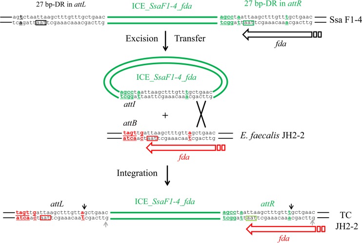
FIG 7.
Localization of the DNA cutting site of integrase by sequencing of the attR site in E. faecalis JH2-2 transconjugants. ICE_SsaF1-4_fda is shown in green in its integrated form in donor S. salivarius strain F1-4 (top), its excised form (middle), and its integrated form in E. faecalis JH2-2 recipient strain after transfer and integration (bottom). Nucleotide differences in att sequences are indicated in green for ICE_SsaF1-4_fda and in red for E. faecalis JH2-2. The position of the DNA cutting site of integrase deduced from this analysis is indicated by a black arrow. Previous work aimed at studying CIME-ICE accretion also identified the same cutting position as well as the position of a second staggered cutting site located 6 bp from the first one (indicated in gray) (48).

