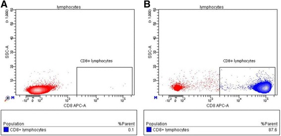
Fig. 3.

Relative content of CD8+ cells in the isolated cultures of HER2-specific T cells (E75-specific cells). The content of CD8+ cytotoxic T cells in isolated fraction was analyzed after culture for 10–14 days in the presence of rhIL-2, rhIL-7, and rhIL-15 stimulants. a The scatter plot shows the events from the lymphocytic region in control sample (unlabeled cells). b Scatter plot showing the events from the lymphocytic region in an experimental sample (anti-CD8-FITC-labeled cells)
