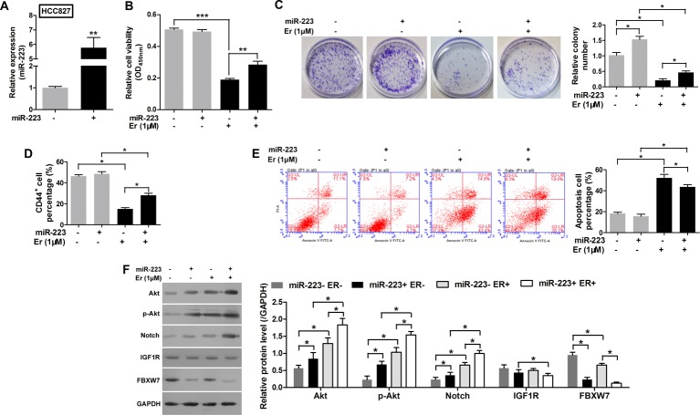
Figure 3. Overexpression of miR-223 reduced the sensitivity of HCC827 cells to erlotinib by enhancing the Notch pathway but not the AKT pathway.
(A) The levels of miR-223 in HCC827 cells transfected with miR-223 mimics or an NC. (B) Cell viability as evaluated with the CCK-8 assay. The HCC827 cells were first transfected with miR-223 mimics and then treated with 1 µM erlotinib. (C) Images recorded with a MicroView imager showing the numbers of colonies formed by HCC827 cells transfected with miR-223 mimics and then treated with erlotinib. (D) The percentage of CD44+ cells in each group of HCC827 cells transfected with miR-223 mimics and then treated with erlotinib. (E) Representative data from FACS analyses of cell apoptosis levels, and the percentage of apoptotic cells among HCC827 cells transfected with miR-223 mimics and then treated with erlotinib. (F) Western blot analyze the expression of p-Akt, Akt, Notch, IGF1R, and FBXW7 in HCC827 cells transfected with miR-223 mimics and then treated with erlotinib. GAPDH served as an internal control. All data represent the mean value ± SD from three independent experiments; *P<0.05, **P<0.01, and ***P<0.001.

