Figure 1.

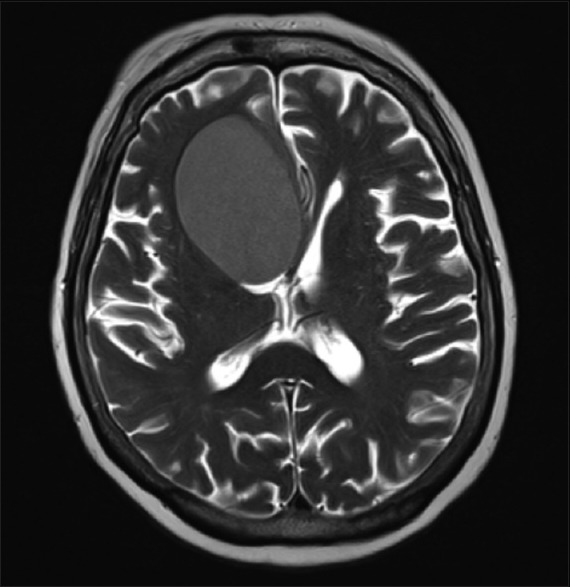
T2-weighted axial magnetic resonance image of the brain demonstrating prominent, hypointense, right cystic craniopharyngioma with mass effect on the right lateral ventricle

T2-weighted axial magnetic resonance image of the brain demonstrating prominent, hypointense, right cystic craniopharyngioma with mass effect on the right lateral ventricle