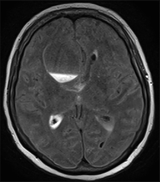
Figure 3.

Fluid-attenuated inversion recovery-weighted axial magnetic resonance image of the brain demonstrating fluid signal within the cystic cavity without change in cyst size and persistent ventricular dilatation, representing a change from the initial imaging findings
