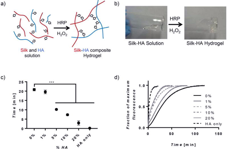
Figure 1. Hydrogel Gelation.

(a) A schematic representing the single step covalent crosslinking between tyrosine residues on silk and tyramine side chains on HA creating a composite hydrogel. (b) Images showing gelation of silk-HA hydrogels during a vial inversion test. (c) Gelation times, as determined by the vial inversion test, show hydrogels consisting of more than 1% HA decreased gelation time (n=4, ***p≤0.001). (d) In addition, the increasing of HA concentration affected crosslinking kinetics by reducing the lag period seen most prominently in 0% hydrogels and decreasing time at which crosslinking was complete (n=7).
