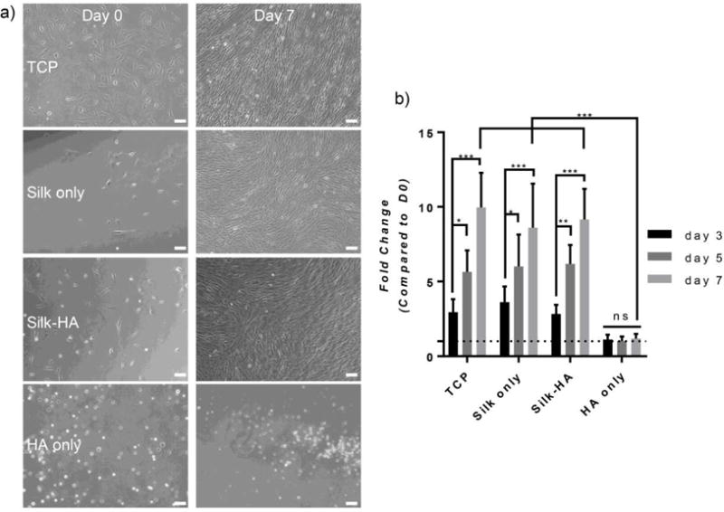
Figure 7. 2D hMSC Response.

(a) Bright-field images of 2D hMSCs are shown at day 0 and 7. Hydrogels containing silk fibroin enhanced cell spreading whereas HA only hydrogels inhibited spreading (scale bar=100 μm). (b) The fold change of hMSC DNA content as compared to day 0 was calculated for days 3, 5, and 7. Silk only and silk-HA (10% HA) hydrogels promoted cellular growth similar to that of TCP whereas HA only hydrogels showed inhibited growth (n=6, * p≤0.05, **p≤0.01, ***p≤0.001).
