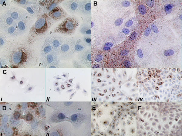
Figure 1.
Immuno-characterization of IEC-18 cell heterotypy. A: Carboxyl IGFBP-2 immunostaining of the sequestered C2 fragment in cells with a preserved crypt cell phenotype but not in others. B: F-actin immunostaining (performed without antigen retrieval to visualize dynamic actin filaments) demonstrates intense staining in crypt cells but not in others. C: Cells plated at variable density with 10% FBS start out as weakly C2 positive (i) but have a progressive loss of C2 immunostaining prior to confluence (ii-iv). D: Immunolocalization of the C2 antigen demonstrates that both C2 positive (i) and C2 negative (ii) cells preserve their phenotypes during proliferation. E-F: Control immunolocalization using the IGF type 2 receptor (E) as a prelysosome-localized antigen and villin (F) as a cytoplasmic-localized antigen to demonstrate that both intravesicular and cytoplasmic antigens can be evenly detected throughout IEC culture.

