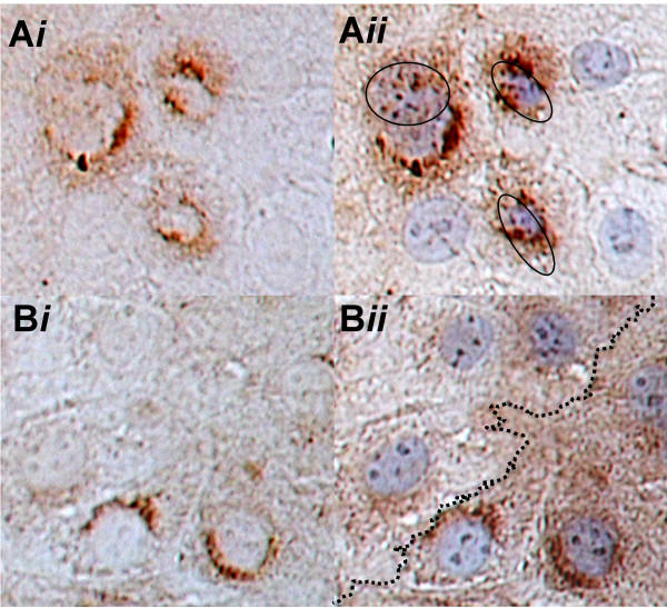
Figure 6.
Double labeling overlay immunolocalization of C2 with either actin or APC. Ai. C2 immunostaining of wet-prepped cells. Aii. C2 immunostaining in the same cells, overlaid with f-actin immunostaining. The proximal cores of actin-stained microvilli bundles located on C2 positive cells are encircled, whereas there are either no or few microvilli on C2 negative cells. Bi. C2 immunostaining of wet-prepped cells. Bii. C2 immunostaining in the same cells, overlaid with APC immunostaining. The dotted line divides C2 positive cells (below) from C2 negative cells (above). There is consistent APC staining in C2 positive cells, whereas there is variable and comparably reduced APC staining in C2 negative cells.

