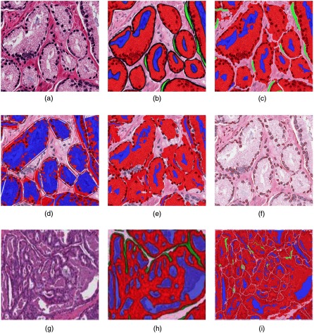
Fig. 9.
We compare various methods in (a)–(f). (a) Original H&E image , (b) annotation image , (c) image by our method, (d) image by Nguyen et al.,23 (e) image by Naik et al.,17 and (f) image by Farjam et al.16 We also illustrate a failure case for our method in (g)–(i). (g) Another input H&E image, (h) corresponding annotation image, overlaid on image in (g), and (i) our result . All the annotation/segmented images are shown overlaid on the corresponding original image and red: gland, blue: lumen, green: periacinar retraction clefting, and white: stroma.

