Figure 2.
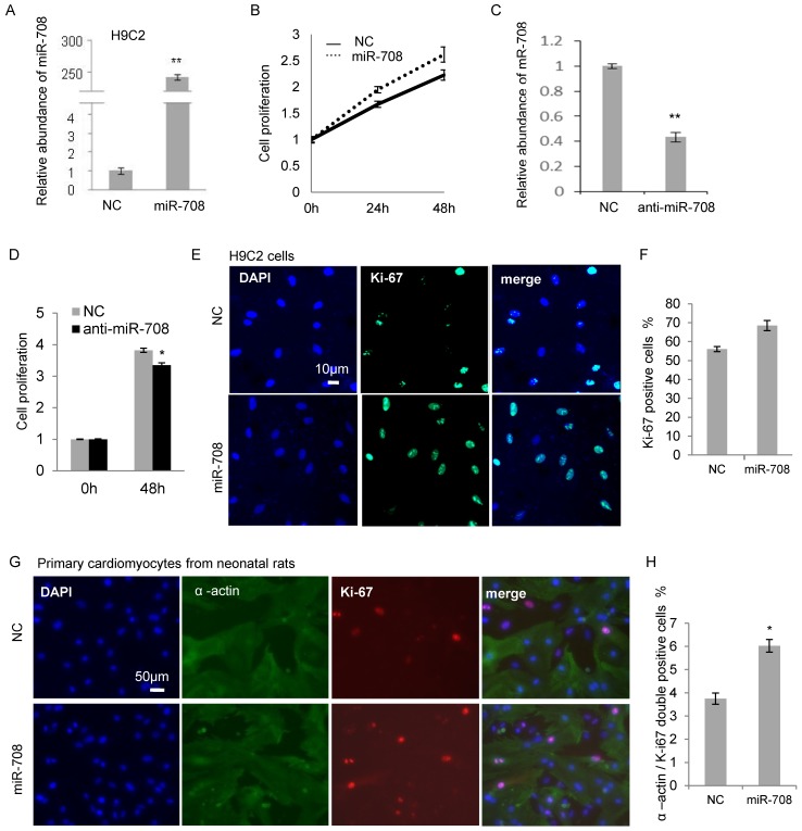
miR-708 promoted the cellular proliferation of cardiomyocytes from neonatal rats. A: Overexpression of miR-708 in H9C2 cells. B: MTT assays demonstrated the increased cell proliferation by miR-708 overexpression in H9C2 cells. C: Knockdown of miR-708 in H9C2 cells by anti-miR-708. D: Decreased cell proliferation by anti-miR-708 in H9C2 cells. E: Ki67 staining indicated a higher Ki67 positive cell proportion in miR-708 overexpressed H9C2 cells. F: Quantitative analysis of Ki67 positive cell proportion in E. G: α-actin and Ki67 staining indicated a higher Ki67 positive cell proportion in the miR-708 overexpressed primary cardiomyocyte cells (α-actin positive) isolated from fresh heart tissue of neonatal rats. H: Quantitative analysis of α-actin / Ki67 double positive cells in G. Data are presented as mean ± SEM (n=3). *p<0.05, **p<0.01.

