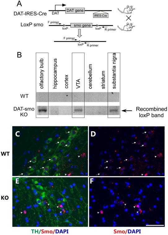
Figure 1. Generation of dopaminergic neuron-specific smo ko mice.

A. Strategy for dopaminergic neuronal specific deletion of the smo gene in DAT-Smo ko mice. B. Detection of recombined alleles (indicating deletion of smo gene) in olfactory bulb, substantia nigra and the ventral tegmental area but not in hippocampus, cortex, cerebellum or striatum of ko mice (lower panel). There is no recombination in different brain areas in wt mice (upper panel). (C) Double immunostaining for TH (green) and Smo (red) shows expression of both Smo protein in TH positive cells (arrow) and TH negative cells (arrow head) in wt mice. (D) shows Smo immunostaining only. However, in ko mice (E) and (F), smo expression is not detected in TH positive cells but is detected in TH negative cells (arrow head). Scale bar= 50um.
