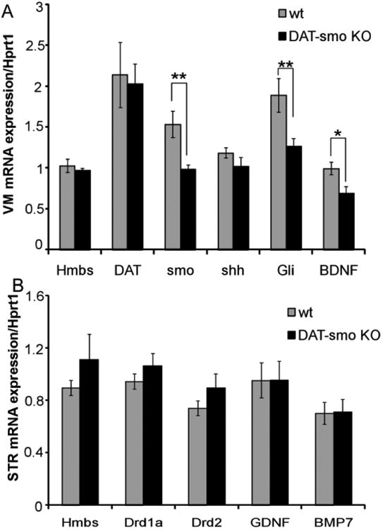
Figure 7. Gene expression alterations in DAT-Smo ko mice after multiple METH exposures.

Gene expression was analyzed using qRT-PCR after 5 days of repeated METH injection in wt and DAT-Smo ko mice. (A) gene expression analysis in ventral midbrain (VM) shows that Smo gene deletion in DA neurons leads to decreased mRNA levels for Smo and target gene Gli in SN and VTA. BDNF mRNA levels are also significantly downregulated in DAT-Smo ko mice (**, P<0.01, * P<0.05, Student's t-test. n= 8). (B) gene expression analysis in striatum (STR) shows no difference in dopamine receptor (Drd1 and Drd2) and neurotrophic factors glial cell line-derived neurotrophic factor (GDNF) and bone morphogenic protein 7 (BMP7) genes, p>0.05 Student's t-test, n=8)
