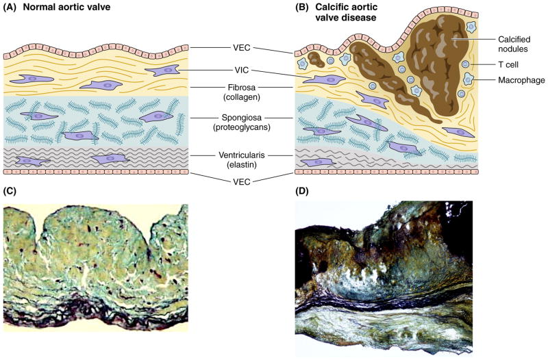
Figure 2.
Structure of a normal and diseased aortic valve. (2A) A healthy aortic valve leaflet contains VECs and quiescent fibroblast-like VICs, and three distinct extracellular matrix layers: a collagen-rich fibrosa, a glycosaminoglycan-enriched spongiosa, and an elastin-rich ventricularis. (2B) Disease progression involves VIC activation, recruitment of immune cells, and subsequent thickening and stiffening of the valve leaflets due to fibrosis and formation of calcific nodules that originate on the fibrosal surface of the leaflet. Histological staining of normal (C) and diseased (D) valve leaflets. Movat’s staining; collagen – yellow; proteoglycans – blue-green; elastin and calcium – black. (C) Modified from Aikawa E. Circulation 2006.2

